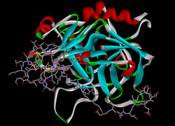
ParM ist ein prokaryotisches Aktin- Homolog [ 1 ] , das die Kraft liefert, um Kopien des R1-Plasmids vor der Zytokinese zu den gegenüberliegenden Enden stäbchenförmiger Bakterien zu treiben .
ParM ist ein Monomer , das in der DNA des R1-Plasmids kodiert und von den Ribosomen der Wirtszelle hergestellt wird . Im Zytoplasma polymerisiert es spontan und bildet kurze Stränge, die entweder an ParR binden oder hydrolysieren . ParR stabilisiert ParM und verhindert dessen Hydrolyse. Sobald ParM an beiden Enden an ParR gebunden ist, lagern sich weitere Monomereinheiten an die Enden des ParM an, und die resultierende Reaktion transportiert die R1-Plasmide zu den gegenüberliegenden Zellpolen . [ 2 ] ParMs verschiedener bakterieller Plasmide können erstaunlich vielfältige helikale Strukturen aus zwei [ 3 ] [ 4 ] oder vier [ 5 ] Strängen bilden, um die korrekte Vererbung des Plasmids zu gewährleisten.
Aktion
In vitro wurde die Polymerisation des ParM-Monomers sowohl mit ATP als auch mit GTP beobachtet . Experimente von Popp et al. deuten jedoch darauf hin, dass die Reaktion GTP bevorzugt und dass GTP das Nukleotid ist, das in der Zelle höchstwahrscheinlich den signifikanten Beitrag leistet. [ 6 ] Im Folgenden wird GTP als das aktive Nukleotid angenommen, obwohl in vielen Experimenten stattdessen ATP verwendet wurde.
ParM bindet und hydrolysiert GTP während seiner Polymerisation . Die vorherrschende Annahme ist, dass eine GTP-„Kappe“ an den Enden der ParM-Polymerstränge erforderlich ist, um deren Hydrolyse zu verhindern. Obwohl GTP nach der Anlagerung durch die ParM-Einheiten hydrolysiert wird, geht man davon aus, dass die Energie, die die Plasmide antreibt, aus der freien Gibbs-Energie der ParM-Monomerkonzentrationen stammt und nicht aus der bei der GTP-Hydrolyse freigesetzten Energie. Die Konzentrationen von ParM-Monomer und -Polymer müssen an den Enden, an denen die Anlagerung erfolgt, im Ungleichgewicht gehalten werden, damit die Reaktion unabhängig von den GTP-Konzentrationen ablaufen kann.
Once the ParM has pushed plasmids to opposite ends of the cell the polymer rapidly depolymerizes—returning the monomer units to the cytoplasm.[7]
Structure
The ParM monomer unit is non-functional before binding a GTP nucleotide. Once the GTP has been bound it can attach to the end of a growing filament. At some point after attachment the ParM hydrolyzes GTP which becomes GDP and remains in the ParM subunit as long as the polymer strand remains intact. ParM forms a left-handed helix structure.[6]
A study by Garner and Campbell has suggested that the unit at the end of the ParM strand must have GTP bound to maintain the stability of the polymer. If one of the ends has the GDP bound version the polymer strand depolymerizes very quickly into its constituent monomer units. This is suggested by their experiment in which they cut growing ParM polymer strands exposing ADP bound ends. Once cut the strands quickly hydrolyzed.[7]
Dynamic Instability
Dynamic instability is described as the switching of a polymer between phases of steady elongation and rapid shortening. This process is essential to the function of eukaryotic microtubules. In ParM, dynamic instability "rescue" or the switch from a shortening phase back to the elongation phase has very rarely been observed, and only when the ATP nucleotide is used. Unbound ParM filaments are found with a typical average length of 1.5 – 2 μm, when the ParM monomer concentrations are 2 μM or more. The dynamic instability of ParM and eukaryotic microtubules is believed to be an example of convergent evolution.[8] L ParM spontaneously forms short polymer segments when it is present in the cytoplasm. These segments serve to very efficiently "search" for the R1 plasmids, and also maintains a favorable concentration of ParM monomer units for polymerization.[6]
References
- ^Gunning PW, Ghoshdastider U, Whitaker S, Popp D, Robinson RC (June 2015). "The evolution of compositionally and functionally distinct actin filaments". Journal of Cell Science. 128 (11): 2009–19. doi:10.1242/jcs.165563. PMID 25788699.
- ^ Hoischen C, Bussiek M, Langowski J, Diekmann S (Februar 2008). „Das Centromer parC des niedrigkopierten Plasmids R1 von Escherichia coli bildet einen U-förmigen Komplex mit seinem Bindungsprotein ParR“ . Nucleic Acids Research . 36 (2): 607–15 . doi : 10.1093/nar/gkm672 . PMC 2241845. PMID 18056157 .
- Popp D, Xu W, Narita A, Brzoska AJ, Skurray RA, Firth N et al. (März 2010). „Struktur und Filamentdynamik des Aktin-ähnlichen Proteins ParM von pSK41: Implikationen für die Plasmid-DNA-Segregation“ . The Journal of Biological Chemistry . 285 (13): 10130–40 . doi : 10.1074/jbc.M109.071613 . PMC 2843175. PMID 20106979 .
- ^ Popp D, Narita A, Ghoshdastider U, Maeda K, Maéda Y, Oda T, et al. (April 2010). „Polymere Strukturen und dynamische Eigenschaften des bakteriellen Aktins AlfA“. Journal of Molecular Biology . 397 (4): 1031–41 . doi : 10.1016/j.jmb.2010.02.010 . PMID 20156449 .
- Popp D, Narita A, Lee LJ, Ghoshdastider U, Xue B, Srinivasan R, et al. (Juni 2012). „Neuartige Aktin-ähnliche Filamentstruktur aus Clostridium tetani“ . The Journal of Biological Chemistry . 287 (25): 21121–9 . doi : 10.1074/jbc.M112.341016 . PMC 3375535. PMID 22514279 .
- Popp D , Narita A, Oda T, Fujisawa T, Matsuo H, Nitanai Y, et al . (Februar 2008). „Molekulare Struktur des ParM - Polymers und der Mechanismus , der zu seiner nukleotidgetriebenen dynamischen Instabilität führt“ . The EMBO Journal . 27 (3): 570–9 . doi : 10.1038/sj.emboj.7601978 . PMC 2241650. PMID 18188150 .
- ^ a b Garner EC, Campbell CS, Weibel DB, Mullins RD (März 2007). „Rekonstitution der DNA-Segregation durch die Assemblierung eines prokaryotischen Aktin-Homologs“ . Science . 315 ( 5816): 1270–4 . Bibcode : 2007Sci...315.1270G . doi : 10.1126/science.1138527 . PMC 2851738. PMID 17332412 .
- ^ Garner EC, Campbell CS, Mullins RD (November 2004). „Dynamische Instabilität in einem DNA-segregierenden prokaryotischen Aktin-Homolog“. Science . 306 ( 5698): 1021–5 . Bibcode : 2004Sci...306.1021G . doi : 10.1126/science.1101313 . PMID 15528442. S2CID 14032209 .